Digital X-Ray Procedures
HSG, IVP, MGU & RGU
USG Guided
Fetal Medicine


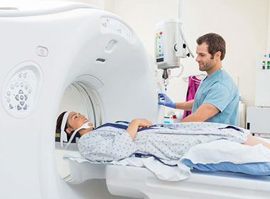
Multi-Slice CT Scanning (MSCT) is a sophisticated imaging method that produces high-resolution, cross-sectional images of the body with remarkable speed and accuracy by utilizing many detector rows. Faster scans and more detailed images are possible with MSCT because it can gather numerous slices in a single rotation, unlike typical CT scans. Numerous medical illnesses, including as pulmonary disorders, malignancies, severe injuries, and cardiovascular diseases, can be diagnosed with this technology. In emergency scenarios, where prompt imaging is essential for prompt medical intervention, MSCT is especially helpful.

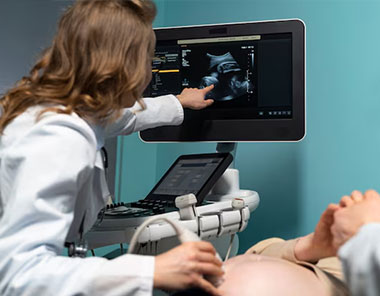
A non-invasive imaging method called an Ultrasound scan produces real-time images of the body's internal structures using high-frequency sound waves. Without using radiation, it is frequently used in medical diagnostics to look at organs, tissues, and blood flow. In addition to cardiology, abdominal imaging, and musculoskeletal evaluations, ultrasound is frequently used in obstetrics to track fetal development. Because the process is quick, painless, and safe, it is a vital tool for early disease detection and treatment planning. Ultrasound now incorporates 3D and Doppler imaging due to technological improvements, which improve diagnostic precision and offer more intricate visualizations.


A non-invasive imaging method called an Ultrasound scan produces real-time images of the body's internal structures using high-frequency sound waves. Without using radiation, it is frequently used in medical diagnostics to look at organs, tissues, and blood flow. In addition to cardiology, abdominal imaging, and musculoskeletal evaluations, ultrasound is frequently used in obstetrics to track fetal development. Because the process is quick, painless, and safe, it is a vital tool for early disease detection and treatment planning. Ultrasound now incorporates 3D and Doppler imaging due to technological improvements, which improve diagnostic precision and offer more intricate visualizations.
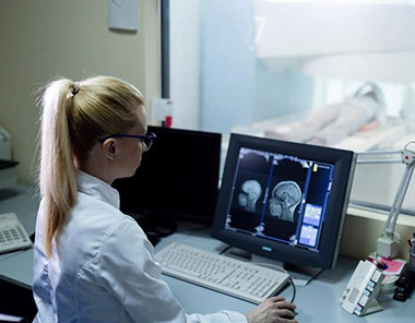
Digital X-rays are a cutting-edge imaging method that produces high-resolution pictures of the body's internal components using digital sensors rather than conventional film. Compared to traditional X-rays, this technology offers quicker results, better image quality, and less radiation exposure. Digital X-rays are frequently used to diagnose lung infections, dental problems, fractures, and other illnesses that impact the soft tissues and bones. Easy storage, sharing, and enhancement of the images for improved analysis can improve patient care and diagnostic precision. Digital X-rays are a vital component of contemporary diagnostic imaging due to their effectiveness, rapidity, and compatibility with electronic medical data.

Our team of committed experts at Aadhaar Diagnostics is committed to provide you professional and considerate diagnostic services. Every stage of your healthcare journey is led by experts who make sure you feel heard, respected, and valued while providing precise, accurate, and dependable outcomes.

